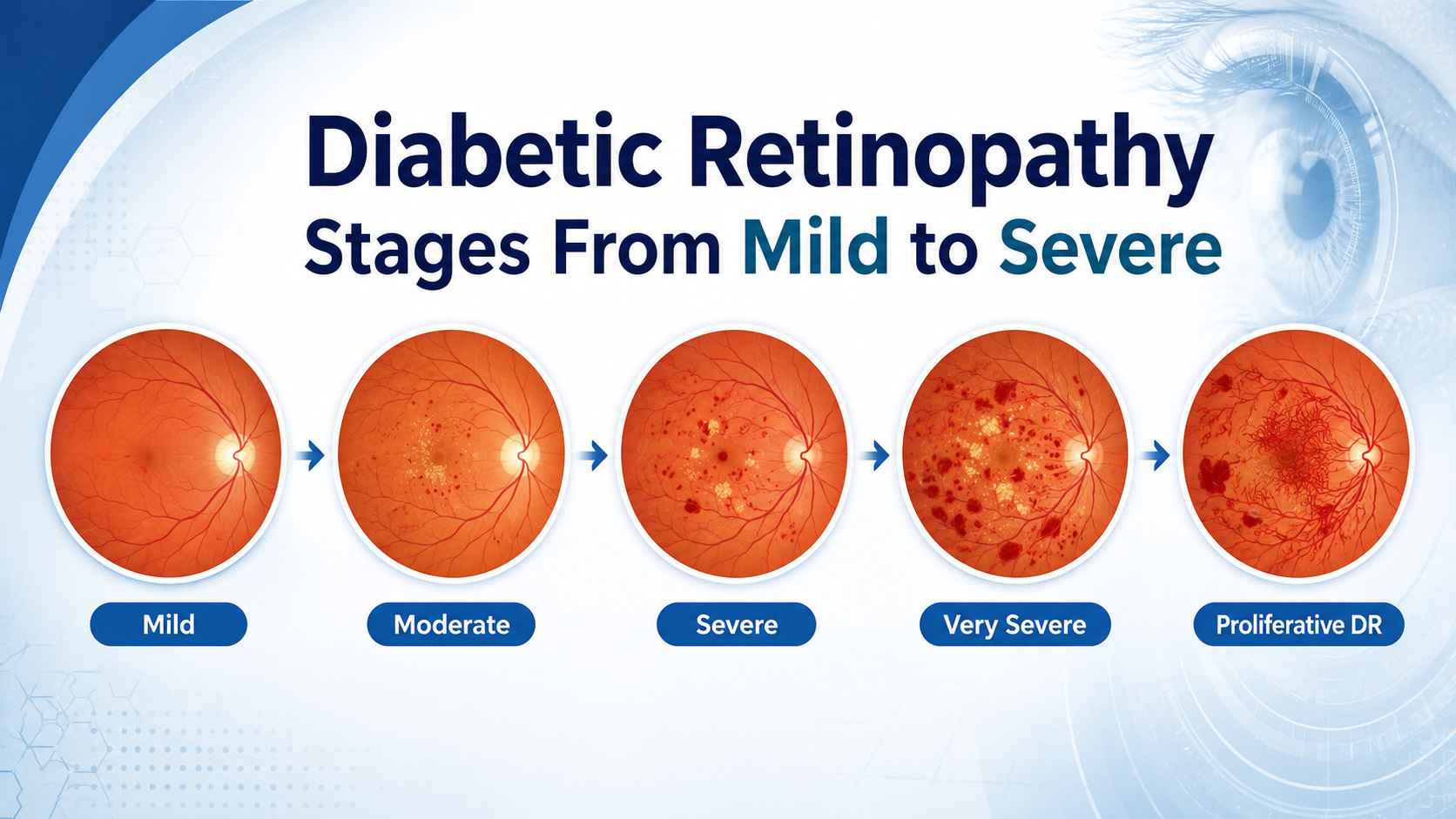
Diabetes can lead to damage of the tiny blood vessels that supply the eye’s light-sensitive tissue (retina). Diabetic retinopathy stages may develop gradually; it is a potential result of these vessels being damaged by persistently high blood sugar. The disease may develop insidiously and may allow the individual to see normally despite the presence of retinal changes.
This is particularly crucial in India. According to the World Health Organization, about 77 million people in India older than 18 years are suffering from type 2 diabetes. It’s also found that over half of those who have diabetes might not be aware of it. Regular screening of the retina is key to preventing unnecessary loss of vision, as diabetes is becoming more prevalent.
Fortunately, early detection, improved diabetes control, and prompt treatment of the eyes can save useful sight. Patients will have a better appreciation of the importance of follow-up appointments when they don’t feel any pain, redness, or blurred vision as a result of the diabetic retinopathy stages.
What Is Diabetic Retinopathy?
Light is turned into signals by the retina, which the brain understands as sight. If blood sugar is high for a long time, the small blood vessels can leak, swell, or get blocked. The eye may start to form abnormal new blood vessels, which are weak and will bleed as the circulation gets worse.
Diabetic retinopathy stage may typically occur in both eyes but may be more severe in one eye than the other. Diseases may occur without any visible symptoms. The Indian Council of Medical Research states that there are no symptoms of diabetic retinopathy in early and late stages. The standard treatment protocol for this includes retinal screening during detection of diabetes and subsequent screening once a year or more often as per the retinal grade.
What Are the Main Diabetic Retinopathy Stages?
There are two general types of Diabetic Retinopathy Stages that doctors classify it as:
Non-proliferative diabetic retinopathy (NPDR): Blood vessels in the retina are damaged, but there are no abnormal blood vessels.
Proliferative diabetic retinopathy (PDR): When the retina lacks sufficient oxygen, the fragile new blood vessels start to grow.
There are three categories of NPDR: mild, moderate, and severe. This results in 4 well-known diabetic retinopathy stages:
- Mild NPDR
- Moderate NPDR
- Severe NPDR
- Proliferative diabetic retinopathy
Diabetic macular edema, also called DME, can develop at any stage. It occurs when leaking blood vessels cause swelling in the macula, the central retinal area responsible for reading, recognizing faces, using a mobile phone, and seeing fine details.
Stage 1: Mild NPDR
Mild NPDR is the first diabetic retinopathy stage that can be seen. Microaneurysms (little balloon-like bulges) occur in the retinal capillaries, which become weakened. This can cause slight leakage of fluid or blood in these areas.
For the majority of patients there is no change in vision. Without a dilated retinal exam, they may keep reading, driving, and working normally, making the disease go undetected.
These changes can be detected and recorded with a fundus exam or retinal photo by an ophthalmologist. If swelling of the macula is suspected, a special test called optical coherence tomography or OCT can give detailed images of the layers of the retina.
Stage 2: Moderate NPDR
Moderate NPDR, is the second diabetic retinopathy stage causing damage to the retina to spread out. There may be an increase in microaneurysms and small retinal hemorrhages. Part of the retina may be deprived of blood supply as some blood vessels leak, swell, or may be partially blocked.
The vision might not yet be affected. But for others, they might experience low or blurred vision, have trouble reading, see spots, or have trouble seeing in low light. These changes shouldn’t be just considered as fatigue, aging, or a showpiece issue.
More careful surveillance is usually necessary at this time. OCT can detect diabetic macular edema, and retinal photography can help the doctor see changes over time. If more information is required regarding leakage, decreased circulation, or blocked vessels, fluorescein angiography or OCT angiography might be recommended. Moderate NPDR does not require injections or laser treatment. The decision depends on whether the macula is affected, whether vision is changing, and how quickly the condition is progressing.
Stage 3: Severe NPDR
An important warning stage is severe NPDR, the third diabetic retinopathy stage. Larger portions of the retina are deprived of oxygen as many blood vessels have become blocked. The retina, in turn, sends chemical messages that stimulate the formation of new blood vessels.
Patients might have very few symptoms, but the risk of developing proliferative diabetic retinopathy is greater. Because severe NPDR is a serious condition, it needs to be closely monitored by a retina specialist, sometimes more frequently than for mild and moderate NPDR.
The ophthalmologist will check for high-risk factors, such as early new vessel formation and bleeding, as well as diabetic macular edema. Close observation may be necessary for some patients, while others may need to receive anti-VEGF injections, laser treatment to the retina, or other treatment. Treatment is individualized, it is difficult to determine from the stage name alone since there is a variation in the involvement of the macula, vision, and systemic health from patient to patient.
Stage 4: Proliferative Diabetic Retinopathy
The most advanced diabetic retinopathy stage is called proliferative diabetic retinopathy. Due to the lack of oxygen to the retina, fragile, abnormal blood vessels form on the retina or optic disc.
They may leak into the clear gel in the eye (vitreous). A small bleed may cause new floaters, dots, or cobweb-like shadows. The larger the hemorrhage, the more sudden and severe the blurring of vision or loss of sight may be.
Scars can also shrink and expand. This may stretch the retina and lead to a tractional retinal detachment. Sometimes, abnormal blood vessels in the front portion of the eye can cause a painful increase in eye pressure.
Patients with diabetes with proliferative diabetic retinopathy need immediate expert attention. Treatment options involve anti-VEGF injection, pan-retinal laser photocoagulation, or vitrectomy surgery when there is some residual bleeding, a lot of scar tissue, or retinal detachment.
How Quickly Can Diabetic Retinopathy Progress?
Progressions are not immediately evident; it takes time. Some do not change over the years, and a few change faster. Factors that affect the risk include the duration of diabetes, blood sugar control, high blood pressure, kidney disease, cholesterol levels, pregnancy, and severity of existing retinal changes.
In India, many people go to the eye hospital only after their glasses have ceased to aid them or they develop blurred vision. By that time, the diabetic retinopathy may have developed. A recent eye exam (which may be a normal vision test) is not a substitute for a dilated retinal examination.
Pregnant women or women who are planning pregnancy and have type 1 or 2 diabetes should talk with their ophthalmologist and obstetrician about retinal screening. It’s important that you have your eyes reviewed during pregnancy, as this condition can develop or worsen.
How Is Diabetic Retinopathy Diagnosed?
A complete diabetic eye examination might involve tests of visual acuity, slit-lamp examination, measurement of eye pressure, and dilated examination of the retina.
Depending on the findings, the doctor may suggest the following:
- Retinal photography to document retinal changes.
- Routine OCT to rule out and quantify macular swelling.
- OCT angiography for evaluation of retinal circulation
- Fluorescence angiography to look for leaks or blockages in the vessels
- Ultrasound B-scan when bleeding or a cataract prevents a clear retinal view
Not all tests are needed for all patients. Investigations are chosen based on symptoms and what is being planned for retinal treatment.
When Should a Person With Diabetes Have an Eye Examination?
Indian guidance recommends retinal screening when diabetes is detected and at least annually thereafter. Patients with varied diabetic retinopathy stage, macular edema, or other risk factors may require more frequent examinations.
An earlier examination may be necessary when:
- Blood sugar has been poorly controlled.
- Blood pressure is high.
- Kidney disease is present
- Pregnancy is being planned
- New visual symptoms have developed.
- The ophthalmologist has already detected retinal changes.
Patients should follow the review date advised by their ophthalmologist instead of waiting until their eyesight changes.
Expert Diabetic Retina Care at Drashti Netralaya
We do it all for you at Drashti Netralaya, from setting you free of cataract surgery cost to dedicated retinal screening and evaluation of OCT/OCTA, laser therapy, intravitreal injections, vitreoretinal surgery, and long-term follow-up if necessary.
Our eye-care team assists individuals with diabetes in knowing what is happening to their vision, determining the current stage, and taking the next appropriate action in a timely fashion.
If you have recently been diagnosed with diabetes, have any changes in your vision or have been referred to a retina specialist, make an appointment for a thorough eye exam. The bottom line for everyone in a different diabetic retinopathy stage is to not wait for blurriness. It is important to be diagnosed early and treated promptly to minimize the potential for permanent vision loss.
FAQ’s
1. Can diabetic retinopathy be present even when vision is normal?
Yes. In the early diabetic retinopathy stage, there are usually no symptoms. It’s possible to read clearly and still have changes in the blood vessels in the retina that need to be monitored.
2. Can better blood sugar control reverse diabetic retinopathy?
Improved blood glucose, blood pressure, and cholesterol control can help to decrease the likelihood of progression; however, once retinal damage is present, it may not go away. Ophthalmoscopy/visual inspection of the retina should be ongoing as recommended.
3. Are spectacles a treatment for diabetic retinopathy?
No. Spectacles correct refractive errors but will not prevent leakage of fluids from the retina, abnormal growth of blood vessels, swelling, and bleeding.